

Conditions: Thyroid Nodule
Thyroid nodules are growths or lumps that form within the thyroid gland. Most thyroid nodules are benign (not cancer). A thyroid nodule may be hyperactive, producing too much thyroid hormone. A thyroid nodule might also grow large enough to compress nearby structures, causing symptoms. Because a minority of nodules are cancerous — commonly estimated at roughly 5 % to 15%, or about 1 in 10—evaluation of a thyroid nodule is undertaken to determine what, if any, treatment is necessary.
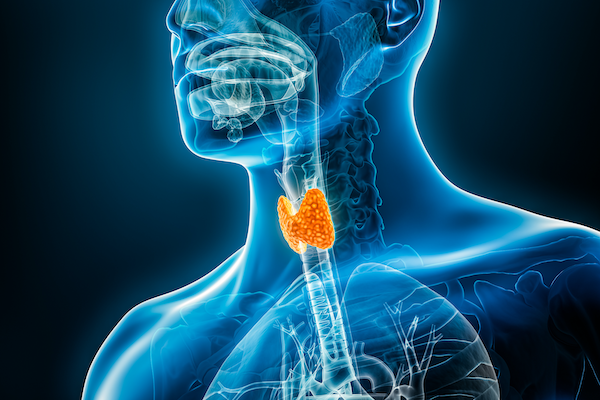

Thyroid gland
About the thyroid gland

Thyroid hormone system. Click to enlarge.
The thyroid gland produces thyroid hormone, which is an essential driver of metabolism and many organ functions. Without adequate thyroid hormone — whether because the gland's production dwindles due to disease, or after surgery or radiation — a hypothyroid state develops. An affected person may become fatigued, gain weight, tolerate cold temperatures poorly, and experience poor concentration or low mood. Too much thyroid hormone — from an overactive gland (or part of it), or from taking too much thyroid hormone medication — leads to a hyperthyroid state with symptoms such as anxiety, jitteriness, tremor, a faster and sometimes irregular heartbeat, and, over time, bone density loss.
Thyroid hormone contains iodine, which must come from the diet. The thyroid is the body's main user of iodine and concentrates it far more avidly than any other tissue, which is what allows radioactive iodine to target the thyroid so specifically. A few other tissues (for example, the salivary glands, the stomach lining, and the breast during breastfeeding) can also take up small amounts of iodine. The thyroid gland sits in the lower neck, draped across the windpipe just above the breastbone (sternum).
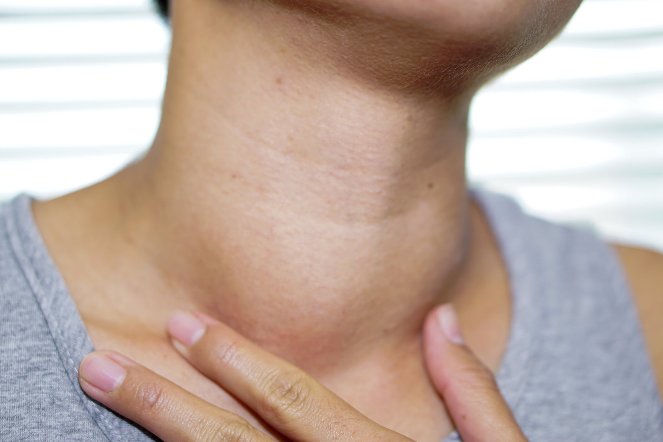

thyroid nodule
The word nodule simply means a lump or an area that looks different from its surroundings. A person may have no nodules, one nodule, or several. Thyroid nodules are very common — they can be found by ultrasound in a large fraction of the general population, and become more common with age. The great majority are benign. Non-cancerous nodules may be of no importance, or they may produce excess thyroid hormone, or occasionally grow large enough to press on nearby structures. Once a thyroid nodule is identified, blood testing (usually starting with a TSH level) and an ultrasound are typically performed.
Thyroid nodules are usually painless, but can occasionally be tender or painful (for example, if there is bleeding into a nodule or inflammation of the gland).
Hyperfunctioning thyroid nodule
A hyperfunctioning thyroid nodule, also known as a toxic adenoma or autonomously functioning nodule, is an area of thyroid tissue that produces thyroid hormone on its own. These make up roughly 5% to 10% of thyroid nodules. Normally, the pituitary gland lowers its output of thyroid-stimulating hormone (TSH) when there is enough thyroid hormone, which in turn tells the thyroid to slow down. A hyperfunctioning nodule ignores this feedback and keeps making hormone regardless, which can lead to hyperthyroidism. A hyperfunctioning nodule is very rarely cancerous. Its location can be confirmed with a radioactive iodine (or technetium) thyroid scan, on which it appears as a "hot" spot. Because these "hot" nodules are almost always benign, they generally do not need a needle biopsy.
Nonfunctioning nodules
Thyroid nodules that do not make excess thyroid hormone are called nonfunctioning ("cold") nodules. These are common and may occur alone or among several nodules in the same gland. Most nonfunctioning nodules are still benign, but as a group they carry a somewhat higher chance of being cancerous than "hot" nodules. For this reason, whether a nonfunctioning nodule needs a biopsy — and ultimately whether it needs surgery — is decided based on its ultrasound features and size, not on the fact that it is nonfunctioning alone.
Thyroid cancer
The word cancer means a growth that has the potential to invade surrounding tissues and spread to other parts of the body, and is therefore more dangerous than a benign (non-spreading) tumor. Most thyroid cancers, when found, are highly treatable.
Once a thyroid nodule is discovered, how is it evaluated?
A thyroid nodule is often found on physical exam or discovered incidentally on imaging (such as a CT, MRI, or ultrasound) done for another reason. When a nodule is identified, a thyroid ultrasound and a blood test of thyroid function (checking for an underactive or overactive thyroid, starting with TSH) are typically performed.

ultrasound
There are many ways thyroid abnormalities may be detected, but the cornerstone method for evaluating them is ultrasound (sonogram). Using only sound waves, ultrasound provides more useful information about thyroid nodules than other imaging types, and it avoids radiation exposure, does not require an IV, and does not involve a magnet or lying inside a narrow tunnel as CT or MRI do. Ultrasound features allow each nodule to be given a standardized risk score. One widely used system, called TI-RADS (Thyroid Imaging Reporting and Data System), scores features such as the nodule's composition, echogenicity (how bright or dark it appears), shape, margins, and any calcifications, and combines them into a level from TR1 (benign) to TR5 (highly suspicious).
The score, together with the nodule's size, guides what happens next:
Some nodules have ultrasound features that reliably indicate they are benign; these may need no needle biopsy and little or no further imaging.
Lower-risk nodules that are below the size threshold for needle biopsy are typically monitored with a repeat ultrasound, often starting around 1 year and continuing at intervals for several years if they remain stable.
Nodules that appear more suspicious for cancer, once they reach the relevant size threshold, are recommended for a needle biopsy.
Importantly, size thresholds for biopsy differ by risk level — more suspicious nodules are biopsied at a smaller size, and nodules smaller than 1 cm are generally not biopsied unless there are unusual concerning features. Needle biopsies are done with ultrasound guidance to confirm accurate needle placement.

Needle biopsy
A thyroid ultrasound usually provides the information needed to decide whether a needle biopsy is warranted, as noted above. A fine-needle aspiration (FNA) is a safe way to obtain a small sample of cells from a nodule for laboratory examination. It is performed with ultrasound guidance to precisely position a very thin needle in the nodule of concern. Only a very small sampling of cells is obtained. Although the needle movements differ from a blood draw or IV placement, the overall risk is low and broadly comparable — some discomfort and possible bruising may occur. While no one likes needles, the risk of an FNA is very low and the information gained is valuable.
An FNA result usually helps determine whether surgery, monitoring, or additional testing is the best next step. The Bethesda System is a well-established way of reporting thyroid FNA results, sorting them into six categories — from benign, through several "indeterminate" categories, to suspicious or malignant — each linked to an estimated chance of cancer and a recommended course of action.
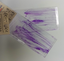


Genetic testing of a thyroid nodule
Examining the cells from an FNA under the microscope often provides enough information to know, with reasonable certainty, whether a nodule is benign or cancerous. When a nodule is clearly cancerous or highly suspicious, surgery is usually recommended. But some FNA results are indeterminate — not clearly benign and not clearly cancerous. This happens especially with follicular lesions, because one type of thyroid cancer, follicular carcinoma, cannot be distinguished from a benign follicular adenoma on a needle biopsy alone; that distinction requires looking at the whole nodule after it is surgically removed.
Traditionally, an indeterminate nodule was handled either by observation with repeat ultrasound and possibly repeat biopsy, or by diagnostic surgery (usually removal of half the thyroid) to get a definitive answer. This meant some patients had surgery only to find out the nodule was benign.
Molecular (genetic) testing was developed to help with exactly this situation. These tests analyze the cells from the FNA sample for genetic changes associated with cancer, in order to refine the estimate of cancer risk for an indeterminate nodule. Unlike the earliest-generation tests, today's molecular tests can meaningfully change management for many patients:
A benign / negative molecular result now carries a low residual chance of cancer (in the range of roughly 3% to 4% with the current tests), similar to a benign biopsy result. This can allow many patients to safely avoid diagnostic surgery and instead be monitored. In studies, roughly half of patients with indeterminate nodules were able to avoid surgery based on these results.
A suspicious / positive molecular result raises the estimated chance of cancer (often to roughly 50% to 65%, and higher with certain specific high-risk gene changes), which supports proceeding with surgery.
No test is perfect. A positive result does not guarantee cancer, and rare cancers can be missed, so a nodule that is not removed still needs ongoing monitoring by a physician. These tests can also involve significant out-of-pocket cost. The main molecular tests currently available in the United States are Afirma (Genomic Sequencing Classifier), ThyroSeq (v3), and ThyGeNEXT/ThyraMIR. (Earlier versions — such as the original Afirma gene expression classifier and ThyGenX/ThyraMIR — and the RosettaGX Reveal test have largely been replaced or discontinued.)

Suggestions for preparing for an appointment are on the Make the Most of Your Thyroid Appointment page.

This page