

Conditions: Head and Neck Tumors (Growths, Lumps, Masses)
Tumors occurring in the mouth, throat, neck, or on the skin may be benign (noncancerous) or malignant (cancerous), and either possibility understandably causes worry. A thorough head and neck evaluation by a head and neck specialist—which may include examining the nose and throat with a thin, flexible scope—along with a possible biopsy are the usual first steps toward a diagnosis. Once a diagnosis is established, the various treatment options, whether surgical or nonsurgical, can be reviewed and discussed. It is worth emphasizing that most neck masses are benign; in adults referred for evaluation, roughly 95% turn out to be noncancerous.
Neck mass
Because of the wide variety of tissue types packed into the small space of the neck, the identity of a lump, fullness, or growth is not always immediately obvious. Certain features of the history and physical examination are very helpful in deciding how to further evaluate and manage a neck mass. In general, neck masses are grouped by how quickly they appear: acute masses (over days to weeks) are most often infectious, subacute masses (over weeks to months) raise more concern for cancer, and chronic masses (over months to years) are frequently congenital or thyroid-related. In adults, a mass that persists for about two weeks or longer without signs of improvement, or a mass whose duration is unknown, is considered concerning for malignancy and generally merits evaluation. Features that further raise concern include a firm mass, one that is fixed to surrounding tissue, one larger than about 1.5 cm, or ulceration of the overlying skin. Evaluation commonly includes a targeted examination of the mouth, throat, and voice box, imaging (often contrast-enhanced CT for non-thyroid masses and ultrasound for thyroid masses), and, when needed, a needle biopsy.

Lymph node enlargement
Enlargement of a lymph node in the neck may simply reflect the node doing its normal job of fighting infection (reactive inflammation). However, persistence, lack of pain, and progressive growth over weeks or months are features that call for evaluation for a more serious problem, such as cancer. A common task for the physician is distinguishing among a node that is enlarged while normally fighting germs, a node enlarged because of a disorder of its function (such as a granulomatous disease like sarcoidosis), and a node that contains cancer cells. When a node without a clear infectious explanation persists—generally beyond about two weeks in an adult, or up to about six weeks after a viral upper respiratory infection—further evaluation may be warranted, such as targeted examination, imaging, and needle biopsy. Cancer within one or more lymph nodes may have spread from a primary cancer elsewhere (for example, tonsil cancer spreading to neck nodes), or may represent a cancer that begins in the lymph tissue itself (lymphoma).
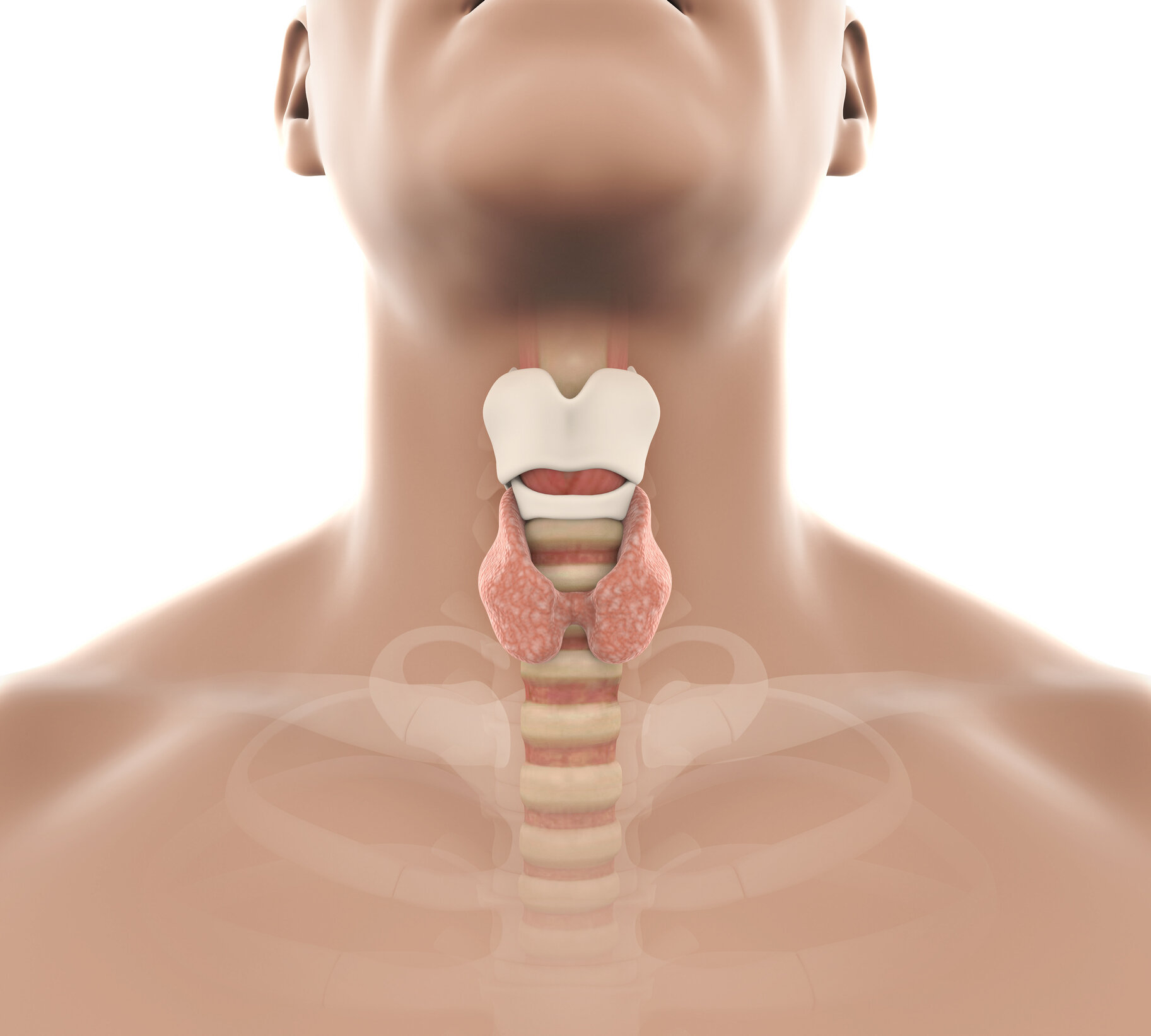

thyroid enlargement and nodules
Located in the lower midline of the neck, the thyroid gland commonly develops lumps referred to as nodules, which are very common in the general population and increase with age. The reassuring news is that the great majority of nodules are benign; only a small fraction are cancerous, with figures commonly cited in the range of about 5% to 15%. The usual workup involves sorting out which nodules have features concerning enough to merit a biopsy—typically guided by ultrasound characteristics and nodule size—and then, based on those results, determining which nodules warrant surgery. Thyroid ultrasound is the standard imaging test, and fine-needle aspiration is the standard biopsy method; when biopsy results are indeterminate, molecular testing can help refine the risk. The main categories of thyroid cancer are papillary thyroid carcinoma (by far the most common), follicular thyroid carcinoma, medullary thyroid carcinoma, and anaplastic thyroid carcinoma. Papillary and follicular cancers are generally well-differentiated and carry a favorable outlook, whereas anaplastic thyroid carcinoma is rare and aggressive. Medullary thyroid carcinoma arises from a different cell type and can be hereditary in a portion of cases.
Parathyroid adenomas and hyperparathyroidism

The parathyroid glands—typically four small glands near the thyroid, but entirely different in function from it—produce parathyroid hormone, which circulates in the blood and regulates the blood calcium level. When one or more parathyroid glands becomes overactive and no longer adjusts hormone production according to the blood calcium, the result is a condition known as primary hyperparathyroidism. This is most often caused by a single benign overgrowth called a parathyroid adenoma. The condition can be insidious, sometimes discovered only through blood tests showing elevated calcium, and over time it may affect the bones, kidneys, and other systems.
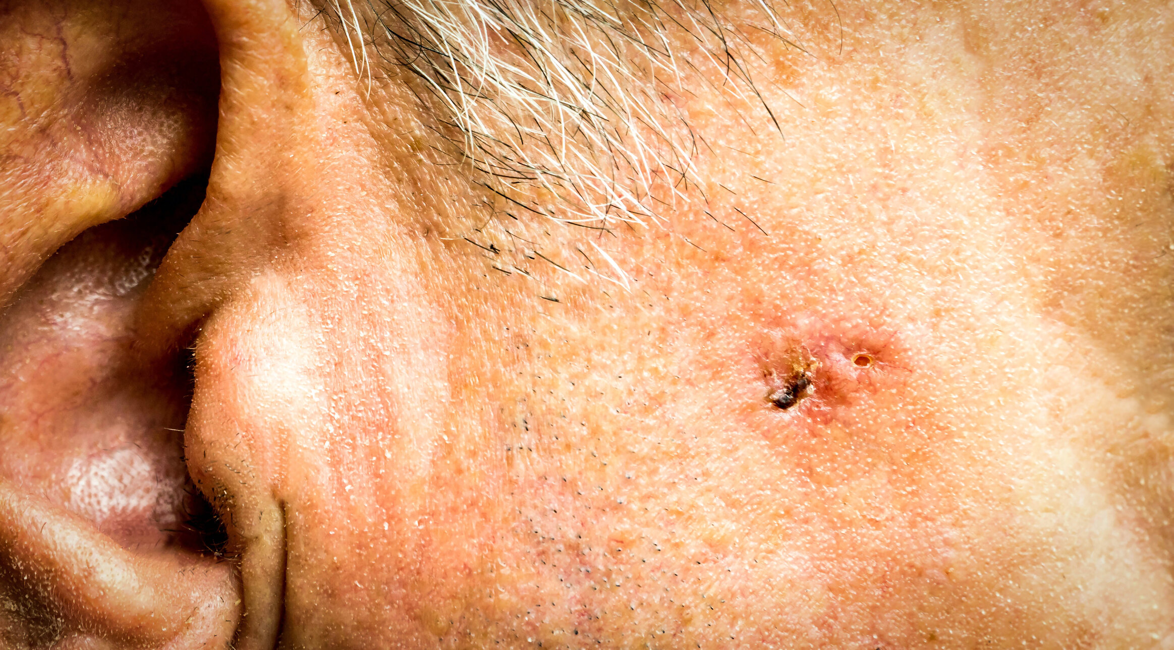

Skin growths
Skin swellings or growths have many causes, both benign and cancerous. Evaluation of new or changing skin growths is how the concerning ones are distinguished from the harmless. Skin cancer is very common—in fact, it is the most common of all cancers. Most skin cancers can be treated effectively, often cured, with surgery to remove the cancer, though other treatments are used in certain circumstances. The specific type of skin cancer generally predicts its behavior, and this information helps guide treatment. Examples of skin lesions and skin cancers include actinic keratosis (a precancerous change), keratoacanthoma, basal cell carcinoma, squamous cell carcinoma, melanoma, and Merkel cell carcinoma.
Oral (mouth) tumors


The mouth includes the lips, the teeth, and the lining surfaces of the oral cavity. Apart from the teeth, the surface of the mouth—called mucosa—is a protective covering that contains glands producing saliva and mucus. Tumors arising from the mucosal surface include both cancers and noncancerous growths. Among the cancers, squamous cell carcinoma is the most common, followed by cancers of the salivary glands (such as adenocarcinoma), and less commonly melanoma. Noncancerous growths include such things as blocked salivary glands (mucoceles), slow-growing bony prominences called tori, viral growths (papillomas), and scar or healing tissue. The tissues surrounding the roots of teeth can also give rise to growths, both cancerous and noncancerous. Tobacco and alcohol use are important risk factors for oral cancers.
Throat (pharynx) tumors

The throat (pharynx) extends from the back of the nose down to the top of the esophagus. This passageway contains several distinct regions, each with different tissue types, which accounts for different tendencies to develop particular cancers. The behavior of a throat cancer depends heavily on its specific type or subtype. For example, squamous cell carcinoma of the tonsil that is associated with the human papillomavirus (HPV)—commonly identified by a marker called p16—behaves differently, and generally carries a more favorable outlook, than a similar tonsil cancer that is not HPV-related. HPV-related cancers of the tonsil and base of tongue have become a leading cause of head and neck cancer, and they can first appear as an enlarging, sometimes cystic, lymph node in the neck. As another example, cancer of the nasopharynx (at the very top of the throat, behind the nose) behaves and is treated differently from cancer of a tonsil.
larynx (voice box) tumors

Growths involving the voice box (larynx) range from areas of swelling (such as vocal nodules or vocal cord edema) to papillomas and other benign tumors, to malignant (cancerous) tumors. Because the sound produced by the vocal cords can be altered by even small changes in their surface, a growth on the vocal cords themselves often causes hoarseness early. In other parts of the larynx, a growth may become quite large before it produces symptoms such as difficulty breathing or swallowing. For these reasons, hoarseness lasting longer than about one month, difficulty swallowing, or symptoms of airway blockage are typically evaluated by a specialist, often including office laryngoscopy—a safe, quick examination of the throat and larynx with a small scope. Further evaluation may include biopsy or imaging, such as a neck CT scan. Some of the more common growths of the larynx include squamous papilloma, vocal nodules, and squamous cell carcinoma. Tobacco and alcohol use are major risk factors for laryngeal cancer.
branchial cleft cyst
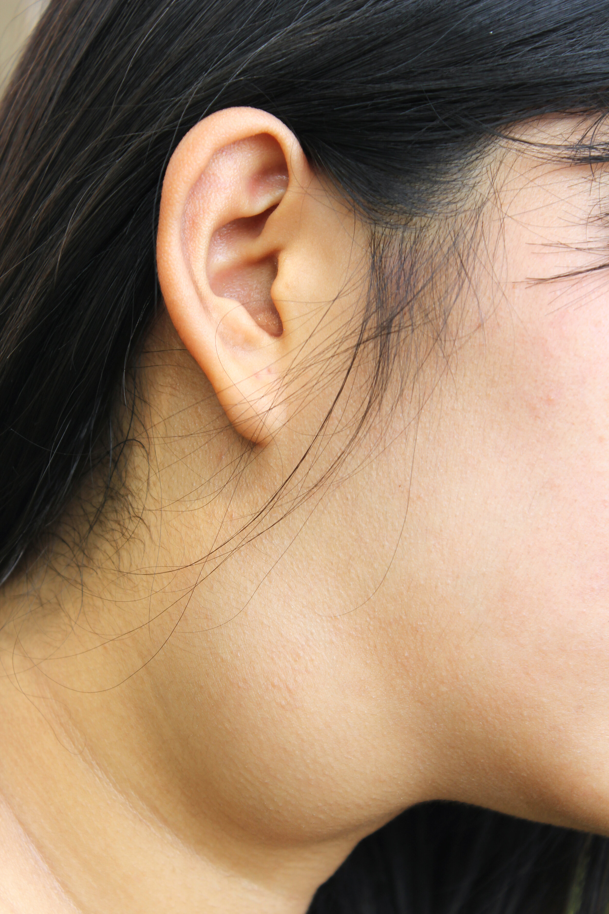
A branchial cleft cyst is a benign condition that can become apparent at any time, can become infected, and requires a definitive diagnosis to distinguish it from cancer. It is a fluid-filled swelling in the upper, lateral part of the neck. It may—but usually does not—have an opening to the skin surface (a sinus or fistula). Branchial cleft anomalies are the second most common congenital neck lesion in children, after thyroglossal duct cysts, and the great majority arise from the second branchial cleft. Many present in later childhood or young adulthood as a solitary, painless mass that was previously unnoticed and has now become tender or enlarged, often after an upper respiratory infection. A branchial cleft cyst is a remnant of embryonic development, resulting from incomplete closure of one of the branchial (pharyngeal) clefts—structures that in fish develop into gills. The diagnosis is often suggested by the history and physical examination because of the relatively consistent location, typically just deep to the front edge of the sternocleidomastoid muscle. Importantly, when a cystic neck mass appears in an adult—particularly over age 40—cancer must be excluded first, because carcinomas of the tonsil, base of tongue, and thyroid can all appear as cystic neck masses. Unlike a thyroglossal duct cyst, a branchial cleft cyst does not typically move up and down with swallowing.
lymphoma

Lymphoma is a cancer of the lymphatic system that can appear in the head and neck but also commonly in the armpits or groin. Persistently enlarged lymph nodes, especially those that are growing and nontender, are suspicious for a tumor. Lymphoma may also be accompanied by fevers, drenching night sweats, or unexplained weight loss. Making the diagnosis generally requires an adequate tissue sample: a surgical (excisional or incisional) biopsy of an entire lymph node is preferred, and a core-needle biopsy is a reasonable alternative when surgery is not practical. Fine-needle aspiration alone is generally insufficient to diagnose and classify lymphoma. The tissue is analyzed using specialized techniques such as examination under the microscope, immunohistochemistry, flow cytometry, and molecular testing to determine the specific subtype, which guides treatment.

Tumor of the parotid salivary gland.

Th three major salivary glands (left to right) sublingual gland, submandibular gland, parotid gland.
salivary gland tumors
There are three paired major salivary glands—the parotid, submandibular, and sublingual glands—along with hundreds of minor salivary glands throughout the mouth and throat. Any of these can give rise to benign or cancerous tumors (neoplasms), which are distinct from swelling caused by infection or a blocked duct. Evaluation of a salivary gland neoplasm often includes fine-needle aspiration biopsy, and imaging (ultrasound, CT, or MRI) may be part of the workup. Treatment depends on the diagnosis but commonly includes observation, surgery, and, in selected cases, radiation therapy. Salivary gland tumors may be described by the specific gland involved or by the type of tumor (based on its appearance under the microscope), which generally predicts its behavior. Features that suggest a benign tumor include a mobile, slowly growing, painless mass with normal facial movement and no associated enlarged neck nodes.

thyroglossal duct cyst
A thyroglossal duct cyst can appear as a lump in the upper neck near the midline, beneath the skin. It is the most common congenital neck mass. Although most present in childhood, they also appear in adults with some frequency. It results from embryonic development: as the thyroid gland normally descends from the base of the tongue down to the lower neck, this pathway occasionally leaves behind a small tract or sac that can later fill with fluid. In other words, the underlying anomaly is present from birth, even though the cyst may not become noticeable until later in life. Characteristically, the mass elevates with tongue protrusion or swallowing because it is attached to the hyoid bone. A thyroglossal duct cyst can become infected, causing pain and swelling. These cysts are benign in more than 99% of cases; in the small fraction that harbor cancer, the type is most often papillary thyroid carcinoma.
others
The list above is far from complete. Other neck tumors include carotid body tumors, glomus tumors, schwannomas (nerve sheath tumors), cancers that have spread to the neck from elsewhere, and many others. After the history and physical examination, biopsy is a mainstay of diagnosis and is often combined with imaging studies to help clarify the nature and extent of the growth.
common questions about lumps, growths, and masses
Finding a lump, growth, or mass in the head or neck is a common reason people see a head and neck surgeon. Most such lumps turn out to be benign (not cancer), but because a minority are serious, they are generally taken seriously and evaluated in an organized way. The questions below explain, in general terms, what these lumps can be and how physicians typically sort them out. This information is educational and does not replace an in-person evaluation.
“I have a lump in my neck, and I do not know what it is.”
Neck lumps have a broad range of causes, and most are not cancer. The most common cause is an enlarged lymph node, often reacting to a recent infection such as a cold, sore throat, or dental problem. Other causes include thyroid nodules or goiter, salivary gland swelling or stones, congenital cysts (such as branchial cleft or thyroglossal duct cysts) that may not be noticed until adulthood, benign fatty growths (lipomas), and, less commonly, cancers that begin in the head and neck or spread to a neck lymph node.
Physicians generally begin with a history and a thorough examination of the head, neck, mouth, and throat, sometimes using a small mirror or a thin flexible camera passed through the nose to view the throat and voice box. Features that tend to be reassuring include a lump that appears suddenly with an infection and then shrinks within two to three weeks, tenderness, softness, and mobility. Features that tend to prompt closer evaluation include a lump that lasts longer than two to three weeks, is hard or fixed in place, is painless and slowly growing, or is accompanied by hoarseness, difficulty or pain with swallowing, ear pain on the same side, unexplained weight loss, or a history of tobacco or heavy alcohol use.
When the cause is not clear from the examination, the usual next steps are imaging (often ultrasound, or a contrast CT scan) and a needle sampling of the lump. A common concept in evaluation is that a neck lump should be followed until it either resolves or receives a diagnosis, because timely evaluation matters when something more serious is present.
“I have a lump in my neck, and the biopsy showed…”
A needle biopsy, called a fine-needle aspiration (FNA), uses a thin needle to remove cells for a pathologist to examine. Results are grouped into general categories, and each category usually points toward a different next stage of evaluation. Common results include:
Reactive or benign lymph node. This means the cells look like an inflamed or normal node, not cancer. It is often reassuring, especially when the lump is shrinking. Because a small number of benign results can miss a problem, a persistent or worrisome lump may still be watched over time or sampled again.
Non-diagnostic or insufficient sample. Not enough usable tissue was obtained to make a diagnosis. This is common and does not itself indicate cancer. Repeating the FNA, often with ultrasound guidance, is a typical next step.
Atypical or indeterminate result. Enough cells were present, but they cannot be classified with confidence. This carries an uncertain risk and usually leads to repeat sampling, a larger biopsy, or additional testing.
Squamous cell carcinoma. This is the most common cancer that spreads to neck nodes and often starts in the throat, mouth, or voice box. When it is found, testing for p16 and HPV (human papillomavirus) is generally performed, because HPV-associated (p16-positive) throat cancers behave differently and tend to have a more favorable outlook. The search then focuses on finding the site where the cancer began.
Thyroid cells or thyroid cancer. This directs evaluation toward the thyroid gland, typically with a dedicated thyroid ultrasound and specialized testing.
Lymphoma. A cancer of the lymph system. Because FNA alone often cannot fully classify lymphoma, a larger core-needle or surgical (excisional) biopsy is usually needed to determine the exact type.
Metastatic skin cancer (such as melanoma or skin squamous cell carcinoma). This points to a skin cancer, sometimes one that was treated or overlooked, and prompts a careful skin examination and staging.
Cystic (fluid-filled) lesion. In younger people this can be a benign congenital cyst, but in adults over 40 a cystic neck node can represent an HPV-related cancer, so these are often sampled again from the solid portion or evaluated with imaging.
Across all categories, results are interpreted together with the examination and imaging rather than in isolation. Biopsy results can take longer than expected when additional specialized testing is required.
“I have a lump, lesion, or growth in my mouth, and I do not know what it is.”
Growths inside the mouth are very common and most are benign. Frequent causes include irritation fibromas (firm bumps from chewing or a rough tooth), mucoceles (mucus-filled cysts of a minor salivary gland, often on the lip), bony outgrowths called tori on the palate or lower jaw, and canker sores or cold sores, which are ulcers rather than true growths. White patches (leukoplakia), red patches (erythroplakia), and mixed red-white patches, as well as lacy white patterns of lichen planus, are also seen. Some white and especially red patches can represent precancerous change, so they receive particular attention.
Evaluation usually starts with a careful look at and feel of the lips, gums, cheeks, tongue (including its sides and underside), floor of the mouth, and palate, with dentures removed. Reassuring features include a clear cause such as a sharp tooth, a lesion that heals within about two weeks once the irritation is removed, softness, and pain that fits an ulcer or infection. Features that generally prompt further evaluation include a sore or lump that does not heal within two to three weeks, a red or mixed red-white patch, firmness or fixation, bleeding, numbness, loose teeth, or a history of tobacco, alcohol, or betel-quid use — though mouth cancer can also occur without these risk factors.
When a lesion is persistent or its cause is uncertain, the usual next step is a biopsy of the tissue to determine whether it is benign, precancerous, or cancerous, which guides what follows.
“I have a lump in my cheek.”
The cheek contains several types of tissue, so a cheek lump can arise from salivary glands (including the parotid gland and small accessory salivary tissue), skin, fat, lymph nodes, nerves, or blood vessels. A common cause is a benign salivary tumor such as a pleomorphic adenoma or Warthin tumor, which typically appears as a slowly growing, painless, rubbery, movable mass. Other causes include salivary gland swelling from infection or a duct stone (often painful and worse with eating), cysts, lipomas, enlarged lymph nodes, and, less often, salivary or skin cancers.
On examination, physicians assess the size, firmness, and mobility of the lump, whether it is painful, and — importantly for the cheek and jaw area — whether the facial nerve is working normally, since this nerve controls facial movement. Reassuring features include slow growth, painlessness, a soft or rubbery feel, mobility, and normal facial movement. Concerning features include rapid growth, pain, a hard or fixed mass, skin changes, or any facial weakness, numbness, or drooping, which raise concern for a more aggressive process.
Imaging is commonly used for cheek and salivary masses, often starting with ultrasound and sometimes adding MRI or CT for deeper or larger lesions. A fine-needle aspiration is frequently performed to help distinguish benign from malignant tissue and to plan any treatment. Because many salivary tumors are ultimately removed surgically, imaging and biopsy also help the surgeon and patient prepare.
“I have a lump in my throat.”
“A lump in the throat” can mean two different things, and they are approached differently. The first is an actual mass that can be seen or felt — for example, enlargement of a tonsil, a growth at the base of the tongue, a thyroid mass pressing on the throat, or a lymph node in the neck. The second is globus: a persistent or intermittent sensation of a lump, tightness, or foreign body in the throat when no actual mass is present. Globus is very common and is usually benign.
Globus has many possible contributors, including acid reflux or laryngopharyngeal reflux, postnasal drip from allergies or sinus problems, tension in the throat muscles, dryness, stress or anxiety, and irritation from smoking or vaping. A helpful reassuring feature of globus is that the sensation often improves rather than worsens while eating and is not usually associated with true difficulty swallowing.
Evaluation generally includes examining the throat and often looking at the voice box with a mirror or a thin flexible camera. Features that typically prompt more testing include actual difficulty or pain with swallowing, food sticking, weight loss, a visible or felt mass, voice change, coughing up blood, or ear pain — findings that point toward a structural cause such as a throat tumor rather than simple globus. When the examination is normal and no warning features are present, the sensation is often attributed to a benign cause; when a mass or a warning symptom is present, imaging, a swallowing evaluation, or endoscopy of the throat and esophagus may follow.
“I have a lump involving the skin.”
Lumps in or just under the skin of the head and neck are extremely common and are usually benign. Frequent causes include epidermoid or sebaceous cysts (which may have a central pore and can become inflamed), lipomas (soft, movable fatty lumps), and other benign growths of hair follicles, sweat glands, or blood vessels. Enlarged lymph nodes can also feel like a skin lump. Less commonly, a skin lump represents a skin cancer such as basal cell carcinoma, squamous cell carcinoma, or melanoma, which is important because the head and neck receive a great deal of sun exposure.
Physicians generally evaluate the lump’s location, size, firmness, mobility, and how long it has been present, along with any changes in the overlying skin. Reassuring features include a soft, movable lump that has been stable for a long time, a cyst with a typical appearance, and a lump that fluctuates with an infection and then settles. Features that tend to prompt further evaluation include a lump that is enlarging, firm and fixed to deeper tissue, larger than a few centimeters, ulcerated or bleeding, or associated with skin discoloration, as well as any pigmented spot that is changing in size, shape, or color.
When a skin lump is stable and typical of a benign cyst or lipoma, it is often simply observed. When the appearance is atypical or concerning, the usual next steps are a skin-focused examination and a biopsy or removal so the tissue can be examined, sometimes with imaging for deeper lumps.

Suggestions for preparing for an appointment are on the Make the Most of Your Head or Neck Mass Appointment page.

This page