Diagnostics: Bethesda System for Reporting Thyroid Cytopathology
What is The Bethesda system?

Needle biopsy is undertaken, usually with ultrasound guidance.
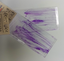
The biopsy specimen is smeared on a glass slide.

The glass slide is sent to a pathologist to examine under a microscope.

This is an example of a fine needle biopsy specimen consisting of only a few cells, but this small sample was adequate for the pathologist to diagnose papillary thyroid carcinoma (Bethesda Category 6).

The appearance of the cells are used to diagnose the specimen. Thyroid nodules are classified according to the Bethesda system.
The Bethesda System for Reporting Thyroid Cytopathology is a standardized system used to categorize the results of fine needle aspiration (FNA) biopsies of the thyroid gland. Developed initially in 2007 during a conference at the National Cancer Institute in Bethesda, Maryland, and most recently updated in 2023, the Bethesda classification system estimates risk of a thyroid nodule being malignant and is used to make treatment recommendations.
The most recently updated Bethesda system (2023) categorizes thyroid FNA results into six diagnostic categories:
Category 1: Non-diagnostic/Unsatisfactory: Samples in this category are inadequate for evaluation and may require repeat FNA or decision making based on other available information. A Bethesda category 1 (Non-diagnostic) score does not show the presence or absence of cancer—it only indicates that no information is available from the fine needle biopsy. Cancer is ultimately found in 13% of nodules initially categorized as Bethesda category 1.
Category 2: Benign: Specimens meeting criteria for this category demonstrate non-cancerous thyroid cells, though still 4% of Bethesda category 2 nodules eventually prove to have cancer. A repeat thyroid ultrasound is usually undertaken after a period of time for these nodules to ensure no significant growth or concerning change occurs. Includes colloid nodules, When cancer is ultimately found in Bethesda category 2 nodules, it is typically a type called incidental micropapillary carcinomas, which means one or more tiny areas of papillary thyroid carcinoma not sampled by the needle biopsy.
Category 3: Atypia of Undetermined Significance (AUS): Samples in this category show some abnormal cells, but it is unclear whether these indicate a benign or malignant process. Subcategories are 1) AUS with nuclear atypia (AUS-N) and 2) AUS with other atypia (AUS-O), with AUS-N carrying a significantly higher risk of cancer (up to 70% in some series) compared to AUS-O. When cancer is ultimately proven by thyroid surgery, nodules in this category are most commonly papillary thyroid carcinoma (often the follicular variant) or Non-invasive Follicular Thyroid Neoplasm with Papillary-like Nuclear Features (NIFTP). Molecular testing is increasingly used to further risk-stratify these nodules.
Category 4: Follicular Neoplasm / Suspicious for Follicular Neoplasm: This category indicates the presence of follicular cells that may suggest a neoplasm, which could either be benign or malignant. Surgical evaluation is usually warranted for a definitive diagnosis, but molecular testing or observation before deciding on surgical management may be used carefully in some cases. Cancers with an initial Bethesda category 4 reading are usually follicular thyroid carcinoma, oncocytic (Hürthle cell) carcinoma, Non-invasive Follicular Thyroid Neoplasm with Papillary-like Nuclear Features (NIFTP) and follicular variant of papillary thyroid carcinoma.
Category 5: Suspicious for Malignancy: This classification suggests a significant concern for thyroid cancer, though it is not definitive. The pathologist may subclassify a category 5 nodule into papillary, medullary, or anaplastic subtypes, which helps guide specific treatment pathways. Over 90% of cancers in this category are papillary thyroid carcinoma, though medullary carcinoma and other types may also be identified. Patients typically undergo surgical intervention for further assessment.
Category 6: Malignant: This category is reserved for samples that clearly exhibit cancerous cells. Initial management typically includes surgical treatment. Cancers in nodules given a Bethesda category 6 are usually papillary thyroid carcinoma, but this category also includes medullary thyroid carcinoma, anaplastic carcinoma, thyroid lymphoma, and cancer from another organ that has spread to the thyroid. Surgery (lobectomy or total thyroidectomy) is the standard approach.
The Bethesda classification system enhances the management of thyroid nodules by providing a framework for assessing the likelihood of cancer and guiding clinical decision-making. It aids in stratifying the risk, which facilitates appropriate surveillance or treatment strategies for patients.
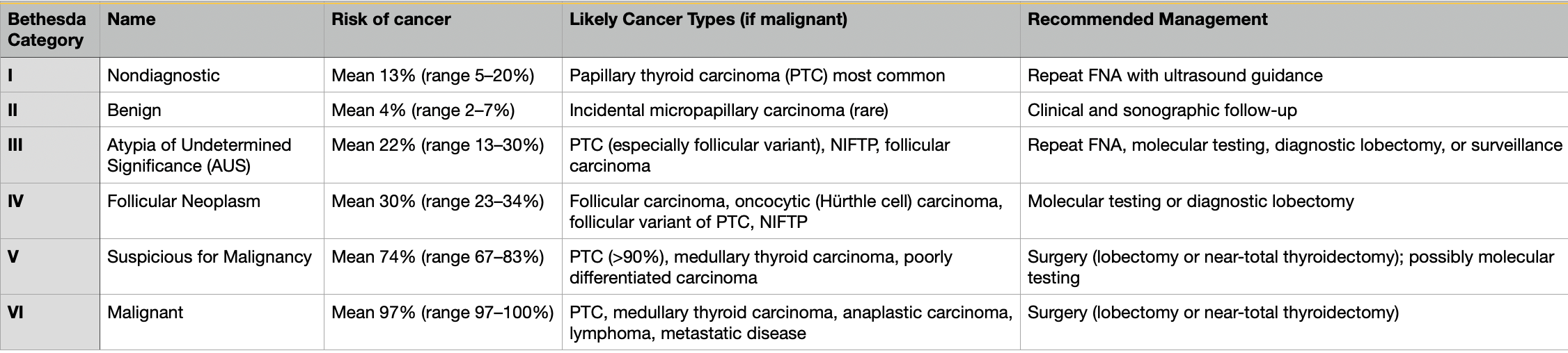
Summary of Bethesda system categories, risk of cancer, type of cancer associated, and the usual recommended management.
