Treatments: Melanoma in the Head & Neck

Wide Local Excision
Wide local excision (WLE) is a surgical procedure used to remove a cancer, with the goal to completely eliminate all cancerous cells at the primary site (where the tumor began). This intentionally includes removing a margin of healthy tissue.
Wide Local Excision for Cutaneous (Skin) Melanoma
For cutaneous melanoma, which occurs on the skin, wide local excision involves the removal of the melanoma along with a margin of normal skin around it. The recommended ideal margin typically depends on the thickness of the melanoma:
Melanoma in situ (involving only the epidermis): a margin of 0.5 to 1 cm
Melanoma less than 1 mm thick: margin of 1 cm
Melanoma > 1 mm up to 2 mm thick: margin of 1-2 cm
Melanoma >2 mm: margin of 2 cm
The procedure aims to ensure that the cancer is not only removed but that there are no residual cancer cells at the edges of the excised tissue, which can be assessed through pathological examination, though these final results are not available until some days after the procedure.
Wide Local Excision for Mucosal Melanoma
Mucosal melanoma occurs in mucous membranes, such as those in the oral cavity, nasal cavity, or other internal surfaces. The approach to wide local excision for mucosal melanoma is similar in that it aims to remove the tumor along with a margin of healthy tissue. However, surgical planning can be more complex due to the anatomical structures involved and potential functional considerations.
Margins for mucosal melanoma are less standardized than for cutaneous melanoma and depend on the location and extent of the tumor. Generally, a margin of at least 1 to 2 cm may be considered, taking into account the need to preserve surrounding structures and functions, such as speech, swallowing, and vision. Final pathological margin status is not available until some days after the procedure.

adjacent tissue transfer (local flaps)
If the area missing skin after removal of a skin cancer is too large to close well by simply stitching the edges together, techniques of borrowing nearby tissue making use of its stretch, skin characteristics, and blood supply are used in local tissue transfer (also called local flaps).
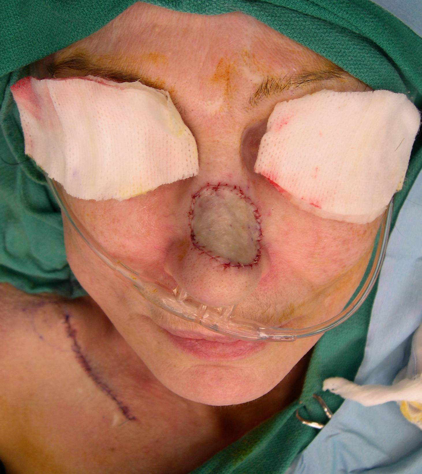
Skin grafts
Another method of reconstructing a skin defect is to use a skin graft, which may be the top layers of skin (a partial thickness skin graft) or the full thickness of skin (a full thickness skin graft). This skin graft is secured under a dressing at the skin cancer site and allowed to heal. The area where the skin graft was taken from is also prepared for healing. At about a week after the procedure, the dressings and sutures are removed. As the skin heals, the color and texture improve.
Lymph node removal (lymphadenectomy or “neck dissection”)
If one or more lymph nodes in the neck have cancer or are at high risk of having cancer spread to them, those lymph nodes typically need to be treated. One method of dealing with these neck lymph nodes is to remove them surgically. This is an effective treatment as well as very informative from a diagnostic standpoint, as the number of lymph nodes with cancer and any adverse features present help to tailor any additional treatments.

Sentinel lymph node biopsy
The sentinel lymph node is the hypothetical first lymph node or group of nodes draining (filtering fluid from) a cancer. It is postulated that the sentinel lymph node is the most likely lymph node to be involved by a cancer if spread has occurred. Therefore, removing and testing a sentinel lymph node can indicate whether spread has occurred, even if too small to be felt on exam or seen on x-rays or other imaging. The sentinel node biopsy is the identification, removal and analysis of the sentinel lymph node (or sentinel lymph nodes if a few good candidates are apparent) of a particular tumor.

External beam radiation (radiation therapy)
Radiation therapy is an effective type of treatment for many cancers, especially small ones. Melanoma is, unfortunately, only partially responsive to radiation therapy, though the techniques of delivering radiation have been adjusted to maximize effectiveness against melanoma.
Radiation therapy may be used after surgery in situations where all other techniques still yield significant risk for recurrence or persistence of the melanoma. Radiation could be applied to the site of the original tumor as well as to areas where the tumor may have spread, such as to the lymph nodes in the area.

Immunotherapy for melanoma
Immunotherapy is a treatment approach that harnesses the body's immune system to combat cancer. This strategy has become increasingly important due to its ability to provide long-lasting responses in many patients, even in later stages of the disease.
One of the primary types of immunotherapy used for melanoma is checkpoint inhibitors. These agents, such as pembrolizumab and nivolumab, work by blocking proteins that inhibit immune responses. By doing so, they allow T-cells to more effectively attack cancer cells. This class of treatment has shown significant efficacy in improving survival rates for patients with advanced melanoma.
Another form of immunotherapy is targeted therapy, which focuses on specific genetic mutations present in some melanoma cells. For instance, patients with BRAF mutations may benefit from targeted agents like vemurafenib or dabrafenib, which can effectively reduce tumor size and improve outcomes.
In addition to checkpoint inhibitors and targeted therapies, vaccines are being explored as a way to enhance the immune response against melanoma. These vaccines aim to stimulate the immune system to recognize and attack melanoma cells specifically, providing a tailored approach to treatment.
Combination therapies are also being investigated, where different immunotherapy agents are used together to improve outcomes. For example, combining a checkpoint inhibitor with targeted therapy or another checkpoint inhibitor may lead to better response rates in patients with advanced disease.
Overall, the use of immunotherapy for melanoma represents a significant advancement in melanoma treatment.

This page