Conditions: Auricular Hematoma and Cauliflower Ear
This page describes auricular hematoma and the potential result, cauliflower ear.

What is an auricular hematoma?
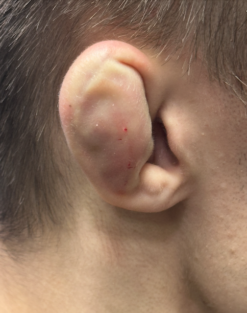
An auricular hematoma is a collection of blood that forms beneath the perichondrium of the ear's pinna (external ear), typically resulting from blunt or shearing trauma to the auricle. It is most commonly seen in contact sports such as wrestling, boxing, martial arts, and judo.
The diagnosis is clinical. The affected ear presents with a fluid-filled, tender swelling over the cartilaginous portion of the auricle, typically appearing shortly after trauma. The earlobe is spared, as it lacks cartilage.
What is the anatomy and physiology of an auricular hematoma?
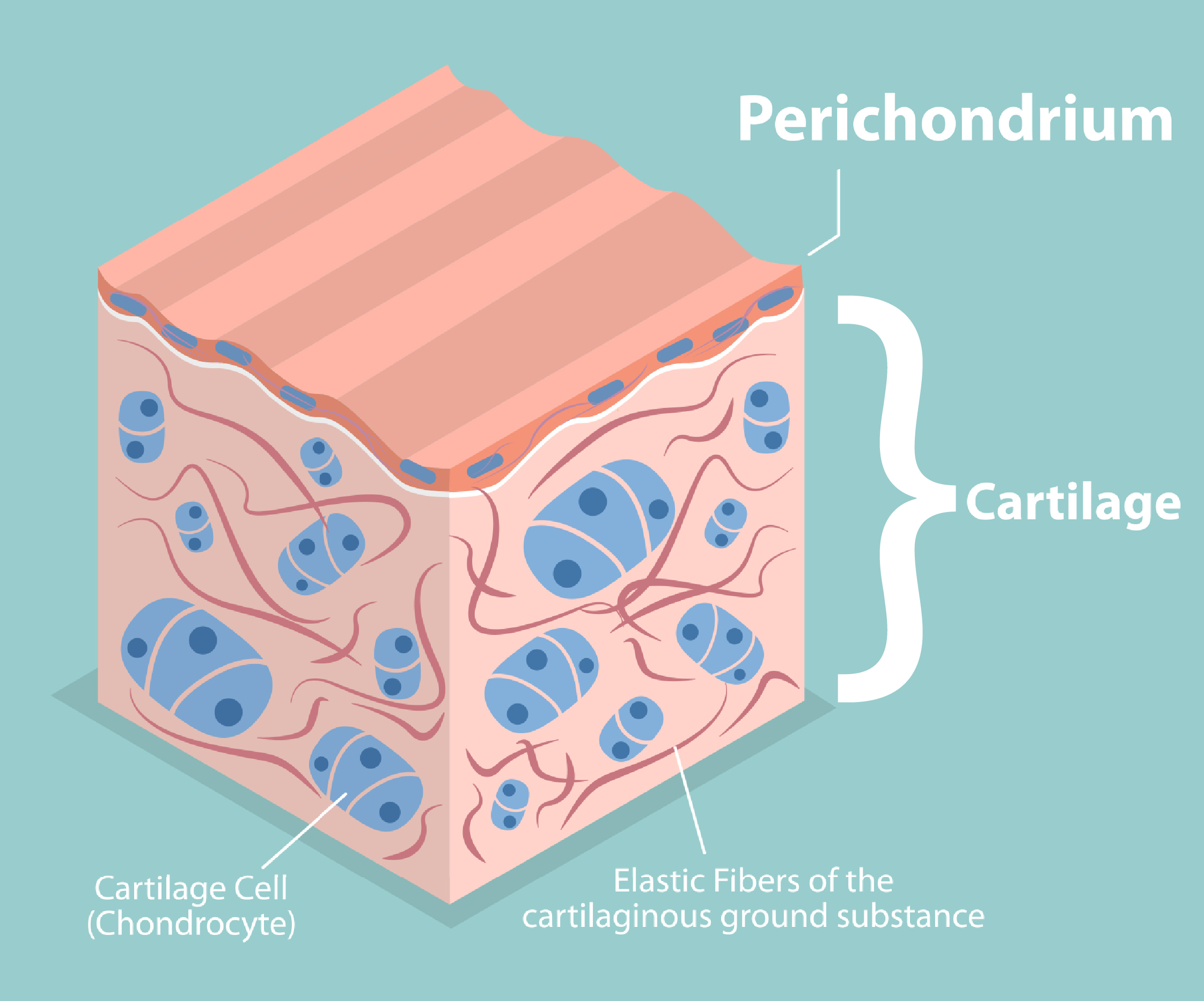
The trauma separates the perichondrium from the underlying auricular cartilage, and blood accumulates as a hematoma in this subperichondrial space. Cartilage has no blood vessels running through it and depends entirely on oxygen and nutrients diffusing from the perichondrium into the cartilage. A hematoma separating the perichondrium from the cartilage prevents oxygen from diffusing into the cartilage. Without treatment, this leads to cartilage death and subsequent scar and new but misshapen cartilage deposition, producing the characteristic permanent deformity known as cauliflower ear (or "wrestler's ear").
How is an auricular hematoma managed?
Drainage is undertaken if a fluctuant hematoma is present within 7 days of injury. Options include needle aspiration or incision and drainage.
Bolster/compression dressing is applied after drainage to prevent reaccumulation of blood in the potential space. Various materials have been used, including dental rolls, cotton bolsters, buttons, silastic sheets, and thermoplastic splints. Application of a bolster dressing is associated with lower recurrence rates.
Antibiotics covering a specific bacterium, Pseudomonas aeruginosa, are typically used for 7–10 days (fluoroquinolones like levofloxacin or ciprofloxacin in adults, amoxicillin-clavulanate in children) to lessen the risk of infection, which could extend to within the cartilage.
Frequent follow-up is recommended, often within 24-48 hours after the first treatment.
Avoidance of trauma, including contact sports for at least 2 weeks.
Note: Delayed or untreated hematomas (beyond ~1 month) generally do not respond to attempts at drainage, as the process has progressed to cauliflower ear.

What is a cauliflower ear?
Cauliflower ear (also called wrestler's ear) is an acquired deformity of the external ear (auricle) caused by one or more auricular hematomas. The hematoma disrupts the perichondrial blood supply to the cartilage. Brief disruption of this blood supply may be tolerated with little or no consequence, but longer disruption without treatment (on the order of days to weeks) leads to loss of cartilage, followed by fibrosis and some new (but misshapen) cartilage formation. This produces the characteristic thickened, irregular, bulbous appearance resembling a cauliflower.
In addition to the permanent cosmetic deformity, functional complications of cauliflower ear include hearing loss and ear wax accumulation due to narrowing of the ear canal and external ear infections.
How is cauliflower ear treated?
Prevention is the best method of avoiding cauliflower ear. Protective headgear during contact sports reduces the risk of auricular trauma.
Acute auricular hematoma should be drained promptly (via aspiration or incision and drainage) to help prevent progression to cauliflower ear. Post-drainage pressure dressings, bolster sutures, or splints are applied to prevent re-accumulation of fluid.
Once established, treatment of a cauliflower ear is difficult and often not undertaken. Surgical treatment attempts may involve excision of the mass of scar and new cartilage using a scalpel or ultrasonic aspirator in an attempt to restore normal ear contour. Success is usually limited.

This page