

Conditions: Subglottic Stenosis and Tracheal Stenosis
what are subglottic stenosis, tracheal stenosis?
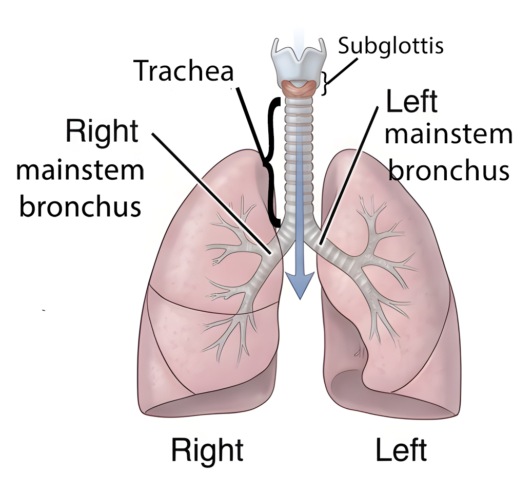
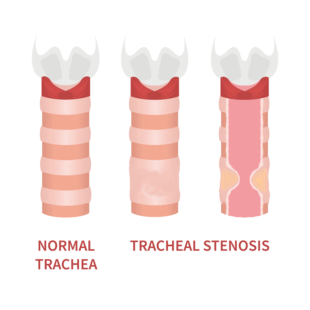
Stenosis means narrowing.
Subglottic means below the vocal cords (glottis).
Subglottic stenosis refers to abnormal narrowing of airway a short distance below the level of the vocal cords.
Laryngeal stenosis refers to abnormal narrowing of the larynx, which is a cartilage framework that houses the vocal cords.
Tracheal stenosis refers to abnormal narrowing of the trachea,
Laryngeaal, subglottic and tracheal stenosis may overlap simultaneously, and the causes and treatments are similar for each. Laryngotracheal stenosis is a narrowing from the vocal cords down to the trachea.
what are the symptoms of subglottic stenosis?
The most common symptom of stenosis of the larynx, subglottis, or trachea is breathlessness (dyspnea) particularly when undertaking physical activities requiring some exertion. Since these types of airway stenosis develop gradually, the symptom of breathlessness typically occur gradually as well. An affected individual may also experience added respiratory sounds (stridor), though mild stridor may sometimes be mistaken for a wheeze. This overlap in presentation between mild stridor and wheezing creates a diagnostic pitfall in which some patients with subglottic stenosis are initially incorrectly diagnosed and treated as having asthma.
what causes subglottic stenosis?
The most common cause of subglottic stenosis (SGS) in adults is prolonged endotracheal intubation, which accounts for the majority of acquired cases. Beyond this, the etiologies can be broadly classified as acquired (far more common) and congenital (rare).
Acquired Causes
• Prolonged or traumatic intubation — This can occur when an endotracheal breathing tube exerts pressure on the subglottic/tracheal lining (mucosa), leading to lack of blood flow, tissue death, then swelling, ulceration and ultimately fibrotic scar formation. Post-intubation subglottic develops in approximately 0.9%–8.3% of intubated neonates, and risk increases with duration of intubation, oversized tubes, multiple intubation attempts, and inadequate sedation. Symptoms typically present 4–6 weeks after the acute injury.
• Idiopathic subglottic stenosis (iSGS) — Accounts for roughly 15%–30% of cases and presents almost exclusively in middle-aged white females. It is a chronic fibroinflammatory condition with no identifiable precipitant. Proposed contributing mechanisms include estrogen-mediated effects, laryngopharyngeal reflux, and aberrant wound healing. Some investigators have suggested iSGS may represent a spectrum of autoimmune-mediated disease.
• Autoimmune and inflammatory diseases — Granulomatosis with polyangiitis (GPA), formerly known as Wegener’s granulomatosis and relapsing polychondritis (RP) are the two most common inflammatory causes. In one multicenter study, GPA accounted for 41% and RP for 26% of non-traumatic, non-tumoral SGS/tracheal stenosis cases. Other inflammatory etiologies include sarcoidosis, amyloidosis, bullous dermatoses, and Crohn's disease.
• Other acquired causes — Tracheostomy and cricothyroidotomy, external laryngeal trauma, chemical or thermal inhalation injury, chondroradionecrosis after radiation therapy, and gastroesophageal reflux disease.
Congenital Causes (present at birth)
Congenital SGS is rare and results from an abnormally small cricoid cartilage. It is the third most common congenital laryngeal anomaly in children.
Key Clinical Considerations
Distinguishing between iSGS and GPA-associated SGS can be diagnostically challenging, particularly when SGS is the presenting symptom of GPA — SGS was the initial disease manifestation in 66% of inflammatory cases in one published series. GPA-related stenoses tend to be circumferential and subglottic, while RP-related stenoses are more often anterior, tracheal, calcified, and extend to the bronchi. Comorbidities such as obesity and diabetes mellitus are frequently observed in patients with acquired SGS and may influence disease severity and recurrence.
how is subglottic stenosis diagnosed?
Clinical Presentation
Patients typically present with progressive dyspnea, noisy breathing (stridor) that occurs with both inhalation and exhalation (biphasic), chronic cough, exercise intolerance, and voice changes. Symptoms depend on the degree of narrowing — mild stenosis (grades 1–2) may be asymptomatic or present only with recurrent upper respiratory infections, while severe stenosis (grades 3–4) causes stridor, respiratory distress, and feeding difficulties in children. The diagnosis is often delayed or considered after other possibilities given its rarity.
Diagnostic Modalities
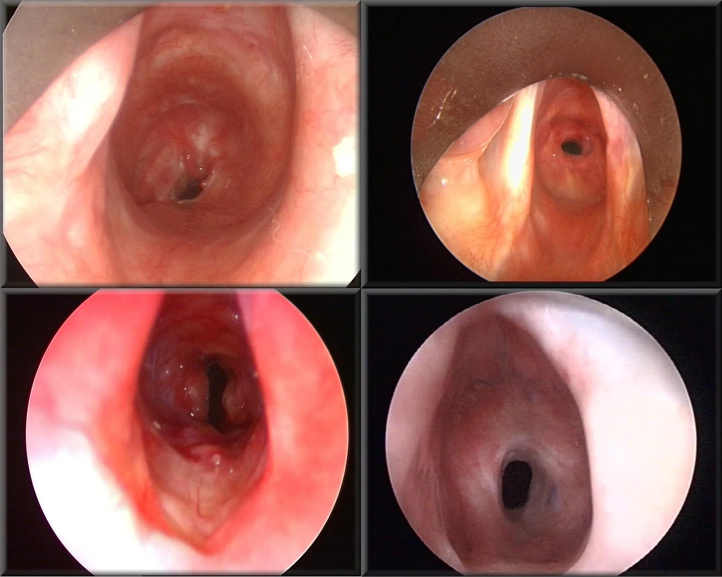
Examples of subglottic stenosis, as viewed from above the vocal cords. The white(ish) structures on the sides of each image are the vocal cords. The narrowing of the airway below this level is visible as the small black passageway. Image Source: Rn cantab, CC BY-SA 3.0 <https://creativecommons.org/licenses/by-sa/3.0>, via Wikimedia Commons
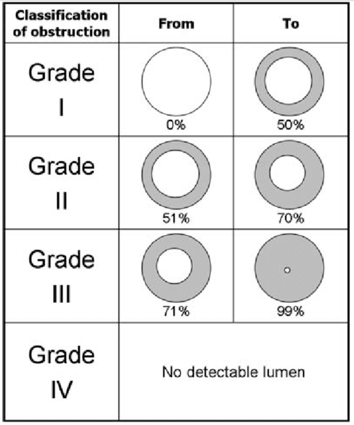
Meyer-Cotton grading scale for severity of subglottic stenosis
Flexible laryngoscopy (awake): Evaluates the upper airway and can sometimes identify subglottic narrowing in the office setting, though the view may be limited.
Direct laryngoscopy and bronchoscopy: The gold standard for definitive diagnosis. Allows direct visualization, grading of stenosis severity, and tissue biopsy when needed. The Myer-Cotton grading scale is the most commonly used classification system, based on the percentage of luminal obstruction: Grade 1 (<50%), Grade 2 (51–70%), Grade 3 (71–99%), Grade 4 (complete obstruction).
CT imaging with multiplanar reconstruction: Highly accurate for detecting laryngotracheal stenosis and provides information on stenosis length, location, and extraluminal disease. Virtual endoscopy can supplement direct visualization. CT can also evaluate airway dynamics during inspiration and expiration.
Pulmonary function testing (PFTs): Useful for surveillance and differentiating upper from lower airway obstruction. The flow-volume loop may show characteristic flattening. The expiratory disproportion index (EDI) has been described as a sensitive and specific tool for differentiating laryngotracheal stenosis from conditions like asthma or COPD.
Laboratory testing: Important to evaluate for autoimmune etiologies such as granulomatosis with polyangiitis (GPA) or relapsing polychondritis, which can cause SGS. ANCA testing and inflammatory markers are usually obtained when an autoimmune cause is suspected.
Biopsy/pathology: Tissue sampling during endoscopy helps distinguish between idiopathic, autoimmune, and other causes.
how is subglottic stenosis treated?
Treatment options, from endoscopic dilation to open airway reconstruction and tracheostomy, are described on the subglottic stenosis treatments page.
Can subglottic stenosis recur after treatment?
Subglottic stenosis can recur after treatment; the risk depends on the cause, the severity and the treatment used, as described on the subglottic stenosis treatments page.

Suggestions for preparing for an appointment are on the Make the Most of Your Throat Appointment page.

This page