

Procedures: Skin Graft
Skin grafting moves healthy skin from one area of the body, the donor site, to cover a wound elsewhere, the recipient site; this page describes how full-thickness and split-thickness grafts are performed and cared for.
What a Skin Graft Is
A skin graft is a piece of healthy skin that is completely removed from one area of the body and moved to cover a wound or defect somewhere else. The area the skin is taken from is called the donor site, and the area it is placed onto is called the recipient site.
A graft is different from a “flap.” A flap is moved along with its own blood vessels still attached, while a skin graft is a thin piece of skin with no blood supply of its own. Because of this, a graft can only survive if it is placed on a wound bed with a good blood supply, which can grow new blood vessels into the graft over the following days.
When Skin Grafting Might Be Used
Skin grafting is a common way to close wounds that are too large or too tight to be stitched closed directly. Situations in which a graft may be used include:
After removal of skin cancer, such as melanoma or other skin malignancies, when the remaining defect is too large to close with stitches
Burns
Large traumatic wounds or injuries
Chronic wounds and ulcers, such as leg ulcers, that do not heal on their own
Areas where the surrounding skin is too tight or scarce to stretch over the wound
The specific reason for a graft depends on the individual situation and is discussed with the surgical team before the procedure.
The Two Main Types of Skin Graft
Skin has two layers: a thin outer layer, the epidermis, and a thicker layer beneath it, the dermis, which contains blood vessels, hair follicles, sweat glands, and oil glands. Skin grafts are classified by how much of these layers they include.
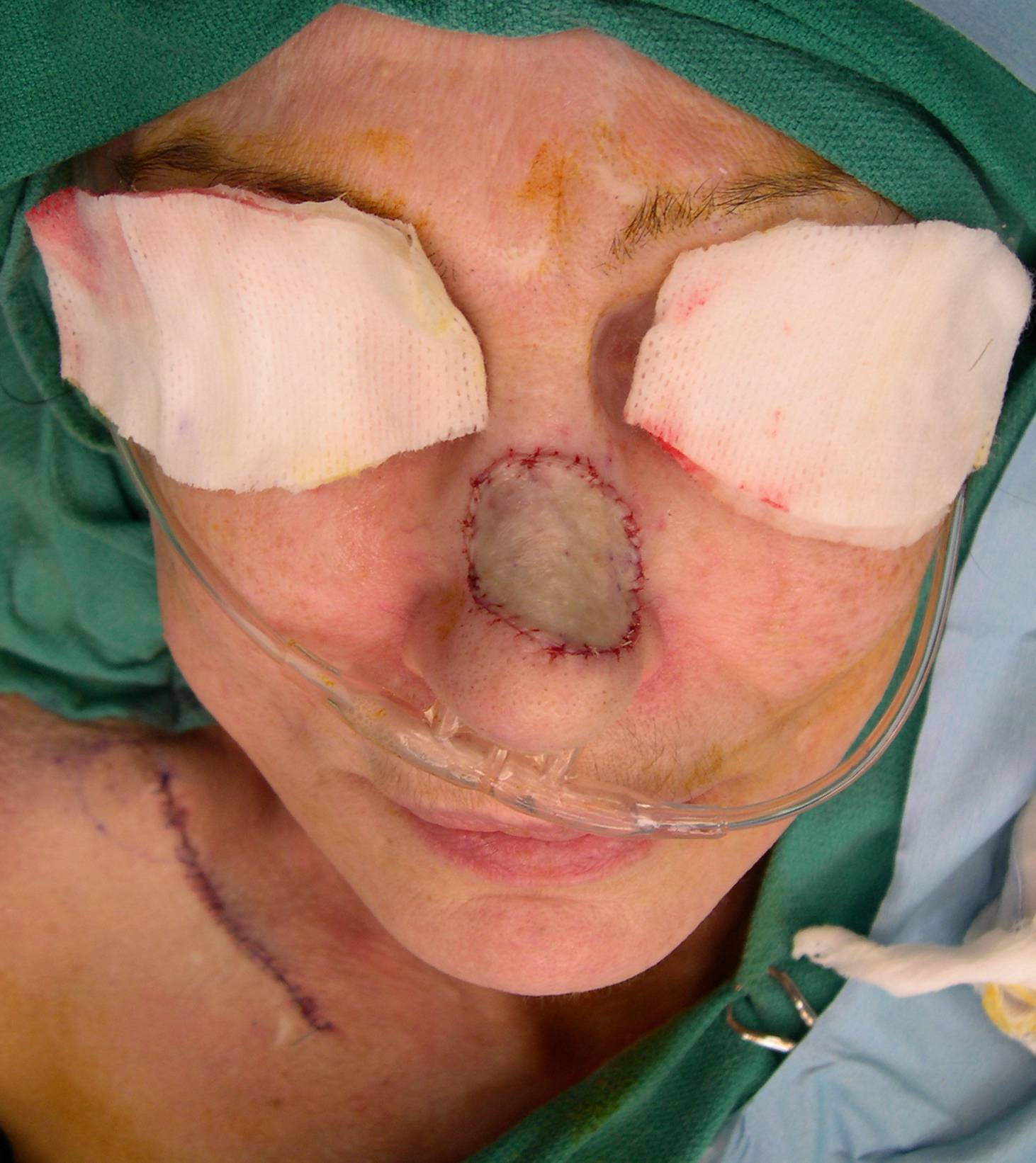
Full thickness skin graft
Includes the epidermis and the entire dermis.
Generally used for smaller areas.
Tends to give a better cosmetic match and shrinks less, which makes it useful for visible or delicate areas such as the face.
Because the whole thickness of skin is removed, the donor site usually needs to be stitched closed, so only areas with some loose, spare skin can be used, such as the groin crease, behind the ear, or the inner arm, often hidden within a natural skin fold.
After the graft is removed, any fat on its underside is trimmed off before it is placed.
Split Thickness Skin Graft
Includes the epidermis and only part of the dermis.
This is the most commonly used type and can cover large areas.
Because only part of the dermis is taken, the donor site heals on its own without stitches.
Split-thickness grafts usually survive reliably, but they tend to look less like normal skin, may differ in color, and can shrink over time.
The donor area is chosen to be easy to reach and hidden by clothing, most commonly the thigh, but also the buttock, upper arm, back, or abdomen. A special instrument, either a powered dermatome or a specialized hand-held knife, removes a thin sheet of skin at a carefully controlled depth; because the deeper part of the dermis is left behind, the donor site regrows its own surface skin from the remaining glands and hair follicles.
Split-thickness grafts are often passed through a device that creates many small slits, turning the graft into a mesh. This lets the graft stretch to cover a larger area and allows blood and fluid to drain out from underneath so the graft can lie flat, with the trade-off of a visible net-like pattern after healing. Grafts for the face are often left un-meshed for a better appearance.
Preparing and Placing the Graft
Before the graft is placed, the wound bed is prepared by removing any dead, damaged, or infected tissue and stopping any bleeding. A clean, healthy, well-supplied wound bed gives the graft the best chance to survive. The graft is then laid over the wound, trimmed to fit, and positioned so it lies flat against the wound bed with no trapped air, blood, or fluid underneath, since trapped fluid is one of the main reasons a graft fails to take.
How the Graft Is Secured
Keeping the graft completely still against the wound bed is essential, because any sliding or shifting can tear the fragile new blood vessels as they try to grow in. Common ways to hold a graft in place include:
Sutures (stitches) or surgical staples around the edges
Fibrin glue, a surgical tissue adhesive, in some cases
A bolster (tie-over) dressing, a bulky pad placed over the graft and tied down with stitches to apply gentle, even pressure and prevent movement
A negative pressure (vacuum) dressing, a sealed sponge dressing connected to gentle suction, which holds the graft down, removes fluid, and can improve graft survival
How a Graft Takes and Heals
A graft survives in stages. For roughly the first two days, the graft has no blood supply yet and absorbs nutrients and oxygen directly from fluid in the wound bed, with a layer of natural fibrin acting like glue to hold it in place. Over the following one to two weeks, new blood vessels grow from the wound bed into the graft and connect with it, restoring a true blood supply; under good conditions, meaningful blood flow is typically established somewhere around five to twelve days. The graft then becomes firmly attached and continues to strengthen and remodel over weeks to months.
Timing depends on location: grafts on the face may reconnect their blood supply in about a week, while grafts on the lower leg may take two weeks or longer. This is why keeping the graft still during this early period is important.
Wound Care and Dressing Changes
The first dressing over the recipient site is usually left undisturbed for a couple of days to a week so the graft can begin to attach. When dressings are changed, they are removed gently, often with counter-pressure on the graft, so it is not accidentally lifted; non-stick dressings are used over the graft, and stitches or staples are commonly removed around a week after surgery.
The split-thickness donor site is often the more uncomfortable of the two wounds, since it is essentially a scrape that must regrow its own surface skin. It is kept covered and moist with dressings such as petroleum gauze, foam, alginate, or specialized moist (hydrogel) dressings, and usually heals on its own within about one to two weeks. A full-thickness donor site is stitched closed and cared for like an ordinary surgical incision, with the stitches removed after a period of healing.
Specific wound care instructions, including when it is safe to get the areas wet or wash them, come from the surgical team and are followed closely.
Recovery and Activity
Protecting the graft from movement by avoiding rubbing, bumping, stretching, or pressure on the grafted area while it is healing
Resting the area, which may include limiting movement, keeping a limb elevated, or avoiding standing and walking for a period if the graft is over a joint or on the leg
A gradual return to normal washing and everyday activity, often around two weeks after surgery if the graft has taken well, though this varies by person and location
Continued change in the color and texture of a graft over many months; grafts may stay drier than normal skin and can be sensitive to sun
Recovery time depends on the size and location of the graft, overall health, and factors such as diabetes and smoking, which can slow healing.
Possible Risks and Complications
As with any surgery, skin grafting carries risks, which can involve either the recipient site or the donor site.
Recipient site: graft failure, in which part or all of the graft does not survive and may need to be repeated; infection, one of the most common reasons a graft fails; collection of blood or fluid under the graft; shifting of the graft from movement or shearing; and scarring, contraction (tightening) of the graft, which can limit movement if over a joint, and a color or texture that does not perfectly match the surrounding skin.
Donor site: pain or discomfort, often more noticeable than at the graft itself for split-thickness grafts; slow healing, infection, or scarring; and lasting differences in skin color or texture at the healed donor area.
Certain factors raise the risk of problems, including infection in the wound, poor circulation, diabetes, smoking, and grafts placed on the lower legs. The surgical team takes steps to lower these risks and monitors healing closely. Increasing pain, redness, warmth, swelling, pus, bleeding, fever, or a dressing that has come loose should be reported promptly to the surgical team.
Key Points
A skin graft moves healthy skin from a donor site to cover a wound at a recipient site, and survives only after new blood vessels grow into it.
Split-thickness grafts include part of the dermis, cover larger areas, and leave a donor site that heals on its own; full-thickness grafts include the entire dermis, give a better cosmetic match, and leave a donor site that is stitched closed.
The graft must be kept still while new blood vessels grow in, a process that mainly takes place over the first one to two weeks.
Both the recipient site and the donor site need careful wound care and monitoring for complications such as infection or graft loss.
This information is general patient education and does not represent a recommendation of specific care. See the site disclaimer for more information, and discuss individual circumstances with a treating physician.

This page