

Conditions: Nasolacrimal Duct
The nasolacrimal duct carries tears from the eyes into the nose, and several distinct conditions from infancy through adulthood can disrupt that drainage system.
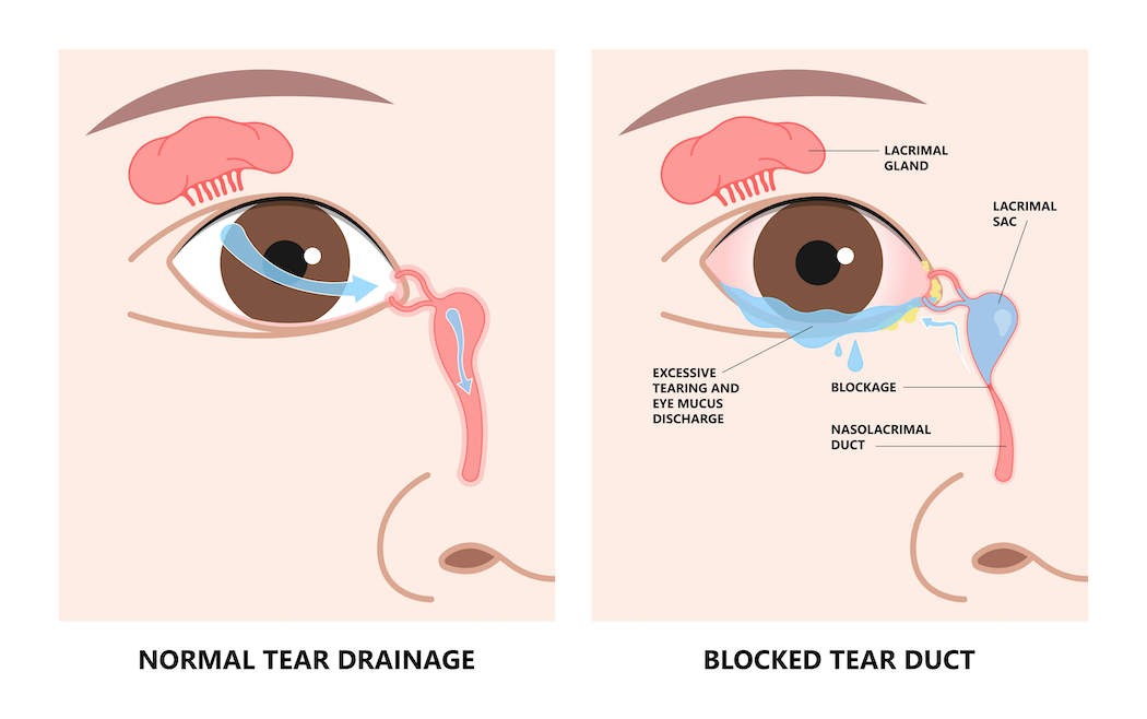
What is the nasolacrimal duct, and how does tear drainage work?
Tears keep the surface of the eye moist and healthy. After washing across the eye, they normally drain through a small system of channels at the inner corner of the eye: tiny openings on the eyelids (the puncta), small connecting channels (the canaliculi), a collecting reservoir (the lacrimal or tear sac), and finally the nasolacrimal duct itself, which carries tears down into the nose. This is why crying or watery eyes often lead to a runny nose as well. When any part of this pathway becomes blocked or narrowed, tears can no longer drain properly and back up onto the surface of the eye and cheek. A blockage can also allow tears and mucus to sit stagnant within the tear sac, creating conditions in which infection can develop.
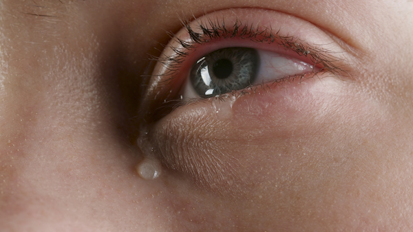
Blocked tear duct in infants (congenital nasolacrimal duct obstruction)
A blocked tear duct is one of the most common eye conditions in infancy, affecting an estimated 5 to 20 percent of babies during the first year of life. In most cases, a thin membrane at the lower end of the duct, where it enters the nose, fails to open normally around the time of birth. Less often, the duct or its drainage openings are narrowed, absent, or did not develop fully, and occasionally trapped fluid swells the tear sac into a cyst called a dacryocystocele, which can appear as a bluish bump beneath the inner corner of the eye. Typical signs begin at birth or within the first few weeks of life and can affect one or both eyes: constant watery eyes, mucus or crusting on the eyelashes (especially after sleep), and recurrent mild discharge or irritation of the surrounding skin. The great majority of blocked tear ducts in infants open on their own, with roughly 90 percent resolving by one year of age.
Blocked or narrowed tear duct in adults (acquired obstruction/stenosis)
In adults, the nasolacrimal duct can become partly narrowed (stenosis) or fully blocked over time, and this is a common cause of watery eyes, occurring most often in middle-aged and older adults, particularly women. Primary, or idiopathic, obstruction has no single identifiable cause, with chronic low-grade inflammation and scarring gradually narrowing the duct. Secondary obstruction follows an identifiable cause, such as infection, chronic sinus disease or a deviated nasal septum, prior nasal or sinus surgery, injury, radiation, certain inflammatory diseases, stones forming within the tear sac, or, rarely, a tumor. Symptoms include persistent watery eyes with tears running down the cheek, sticky discharge or mucus in the corner of the eye, swelling or fullness at the inner corner of the eye, and blurring of vision from the film of excess tears.
Tear sac infection (dacryocystitis) and watery eyes (epiphora)
When tears and mucus become trapped in a blocked tear sac, the stagnant fluid can become infected, a condition called dacryocystitis. It can occur in infants, usually from a congenital blockage, and in adults, usually from an acquired one. Signs include pain, redness, warmth, and swelling at the inner corner of the eye just below the eyelid; a tender lump over the tear sac; discharge of pus; tearing; and sometimes fever. Dacryocystitis can be sudden and severe (acute) or low-grade and long-standing (chronic); new redness, swelling, or pain in this area is generally regarded as a reason for prompt evaluation, since the infection can occasionally spread to nearby tissue. Epiphora, or excessive tearing, is a symptom rather than a single disease, with two broad categories of cause: overproduction of tears in response to irritation, dryness, wind, allergies, infection, or misdirected eyelashes; and impaired drainage, in which the system is blocked or narrowed somewhere along its length, or the eyelid is turned inward or outward so that tears cannot reach the drainage openings. A blocked or narrowed nasolacrimal duct is among the most common causes of epiphora, though in some individuals symptoms occur even when testing shows the drainage passage to be open, a pattern known as functional epiphora, in which the system is patent but pumps tears poorly.
How these conditions are diagnosed
Diagnosis generally begins with a review of symptoms and an examination of the eye and eyelids, including gentle pressure over the tear sac to check whether tears or mucus reflux through the drainage openings. Additional tests may include a dye disappearance test, in which a drop of harmless colored dye is placed in the eye and its persistence after several minutes is used to gauge drainage; tear duct irrigation, in which fluid is gently flushed through the drainage system to identify the location of a blockage; nasal endoscopy, using a small camera to examine the inside of the nose for structural causes; and, in selected cases, imaging studies such as CT, MRI, or dedicated dye-based imaging of the tear drainage pathway. A range of treatment options exists for these conditions, from observation and simple supportive measures to office procedures and surgery, with the approach depending on the specific condition, its cause, and the patient's age.

This page