

Diagnostics: Molecular Testing of a Thyroid Nodule FNA specimen
Molecular testing analyzes the genetic material in a thyroid nodule biopsy sample to help refine the estimated likelihood of cancer when microscope examination alone is inconclusive.
When a thyroid nodule is biopsied and the result is “indeterminate,” meaning neither clearly benign nor clearly cancerous, molecular testing can examine the genetic material in the biopsy sample to better estimate whether the nodule is likely benign or cancerous. This information can inform discussions about whether a nodule may be safely observed or whether surgery is needed, and if so, how much surgery. This page explains what these tests are, how they work, what they can and cannot show, and practical issues such as sample collection, cost, and insurance.
What is molecular testing?
A thyroid nodule is a lump within the thyroid gland, a common finding that is usually benign. When a nodule is evaluated, the first step is often a fine needle aspiration (FNA) biopsy, in which a thin needle collects a small sample of cells that a pathologist examines under a microscope (called cytology).
Molecular testing is an additional laboratory analysis performed on cells collected during that same biopsy. Instead of only looking at how the cells appear under the microscope, molecular testing examines the genes inside the cells for changes associated with thyroid cancer. It does not replace the microscope examination; it adds information to it.
Why is molecular testing used?
For most nodules, the biopsy gives a clear answer: benign (the most common result) or cancerous. In roughly 1 in 5 to 1 in 3 biopsies, however, the microscope findings are “indeterminate,” meaning the appearance is not clearly benign and not clearly cancerous.
Pathologists group biopsy results using a standardized scale called the Bethesda System. Molecular testing is used mainly for the two indeterminate categories:
Bethesda III (atypia of undetermined significance): historically about a 1-in-5 chance of cancer.
Bethesda IV (follicular neoplasm or suspicious for follicular neoplasm): historically about a 1-in-3 chance of cancer.
In these categories, the biopsy alone cannot settle the question, and in the past the only way to know for certain was surgery to remove part of the thyroid. Molecular testing offers a way to refine the estimate of cancer risk without surgery, so that many people with benign nodules may avoid an operation that would not otherwise have been needed. It is sometimes also applied to Bethesda V (“suspicious for malignancy”) nodules, where it is usually used to gather information that may help plan treatment rather than to decide whether cancer is present.
What does the test look at?
Thyroid cancers are driven by specific changes in genes. Molecular tests look for several kinds of changes, including:
DNA mutations: small “spelling errors” in genes such as BRAF, RAS, and TERT.
Gene fusions (rearrangements): two genes that have become abnormally joined together, such as RET or NTRK fusions.
Gene expression patterns: how active thousands of genes are, which can produce a signature that looks more like a benign nodule or more like a cancer.
Different commercial tests emphasize different combinations of these features.
The main tests available
Three molecular tests are most widely used in the United States. They perform similarly overall, though they differ in method and in the additional information they provide.
ThyroSeq v3: a next-generation sequencing test that examines a large panel of thyroid cancer–related genes for mutations, fusions, copy-number changes, and abnormal gene activity. Results are generally reported as negative (benign-appearing, low cancer risk) or positive (a genetic alteration was found, raising cancer risk), and the report often names the specific alteration found.
Afirma Genomic Sequencing Classifier (GSC): an RNA-based test that analyzes gene activity across thousands of genes and reports a result of benign or suspicious. When a nodule is suspicious (or already suspicious or malignant on cytology), an add-on called the Afirma Xpression Atlas can identify specific mutations and fusions to provide additional detail that may inform treatment.
ThyGeNEXT / ThyraMIR: a two-part test that combines a mutation-and-fusion panel (ThyGeNEXT) with a microRNA expression classifier (ThyraMIR) to sort nodules into lower-risk or higher-risk groups.
In broad terms, all three report results along a spectrum from benign-appearing or negative (reassuring) to suspicious or positive (concerning), and the more detailed tests also report which specific genetic changes, if any, were detected.
How is the sample collected?
Molecular testing uses the same kind of needle biopsy already used to evaluate the nodule, often performed with ultrasound guidance. In practice, this usually takes one of two forms:
Extra passes: a few additional needle passes into the nodule during the biopsy collect extra material that is set aside for molecular analysis.
A dedicated collection: the sample is placed into a special preservative tube or collection kit provided by the testing company and shipped to that company’s laboratory.
Often the sample is sent for molecular testing only after the microscope result is found to be indeterminate. Some centers collect and hold extra material at the time of the biopsy so that a second needle procedure can be avoided; others perform a repeat biopsy if molecular material is needed. If a sample is inadequate or degraded, a repeat biopsy may be required.

Needle biopsy is undertaken, usually with ultrasound guidance.
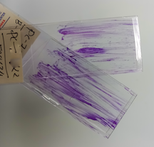
The biopsy specimen is smeared on a glass slide.

The glass slide is sent to a pathologist to examine under a microscope.

This is an example of a fine needle biopsy specimen consisting of only a few cells, but this small sample was adequate for the pathologist to diagnose papillary thyroid carcinoma (Bethesda Category 6).

The appearance of the cells is used to diagnose the specimen. Thyroid nodules are classified according to the Bethesda system.
What the results can and cannot show
Two numbers describe how these tests are interpreted:
Negative predictive value (NPV): how trustworthy a “benign” or “negative” result is. For the major tests, NPV is high (roughly 94–97%). This means a benign result makes cancer unlikely, which is why these tests are best at “ruling out” cancer. Long-term follow-up studies have found that most indeterminate nodules with a benign molecular result remain stable, with very few later found to be cancer.
Positive predictive value (PPV): how trustworthy a “suspicious” or “positive” result is. PPV is more modest and varies by test and by which genetic change is found (often in the range of about 45–75%). This means a positive result raises concern but does not by itself prove cancer, because some nodules with a positive result turn out to be benign.
Important limitations include:
A benign or negative result greatly lowers, but does not completely eliminate, the chance of cancer. Rare cancers are missed.
A suspicious or positive result is not a diagnosis of cancer. Certain changes, such as RAS mutations, are found in both benign and cancerous nodules, making them less specific.
Occasionally the sample is inadequate or degraded, or the result is “not informative,” in which case a repeat biopsy or another approach may be needed.
Molecular results are meant to be interpreted together with the ultrasound appearance (for example, the TI-RADS risk category), the Bethesda cytology findings, and the individual’s overall clinical picture, not in isolation.
The role of specific mutations and fusions
Different genetic changes carry different meanings:
BRAF (especially BRAF V600E): the most common mutation in papillary thyroid cancer. When found, cancer is very likely, and in some cases it is associated with more aggressive features.
RAS: found in some cancers but also in benign nodules, so it is less definitive. RAS-related tumors often behave in a more indolent (slow-growing) manner.
TERT promoter and TP53: less common changes that, when present (particularly alongside BRAF), suggest more aggressive disease.
RET, NTRK, and ALK fusions: abnormally joined genes that can drive cancer. Beyond diagnosis, these are important because FDA-approved targeted drugs exist for certain fusions and mutations (for example, RET inhibitors for RET-driven cancer and NTRK inhibitors for NTRK fusion–positive cancers). This is most relevant for advanced or spreading thyroid cancers, where identifying the driver can open up specific treatment options.
Limitations, cost, and insurance
Not a perfect test: no molecular test is 100% accurate. Both false-negative and false-positive results occur, and results are best viewed alongside the full clinical picture. Much of the published test performance comes from nodules that were surgically removed, so real-world accuracy in nodules that are observed rather than removed is still being studied.
Availability: these tests are performed at specialized central laboratories, and ordering practices vary between larger medical centers and smaller practices.
Cost: the list price of these tests is high, commonly several thousand dollars (often quoted in the range of about $3,000 to $8,000). Actual out-of-pocket costs depend on the insurance plan.
Insurance: coverage varies. Many plans cover molecular testing for indeterminate nodules because avoiding unnecessary surgery can be cost-effective, but prior authorization is sometimes required and coverage is not guaranteed. Expected costs, coverage, and financial-assistance programs are typically addressed by the ordering physician’s office and the testing company.
How molecular results may affect decision-making
Molecular results help match the intensity of treatment to the estimated risk of a nodule. Studies suggest this improves the likelihood that patients receive an appropriate amount of surgery, or avoid surgery entirely. In general:
Observation (surveillance): a benign or negative result, especially with a reassuring ultrasound, is often associated with monitoring the nodule with periodic ultrasounds instead of surgery.
Diagnostic lobectomy (removal of one lobe): an intermediate or positive result may lead to consideration of removing the half of the thyroid containing the nodule, which both establishes a diagnosis and treats a small, non-aggressive cancer.
Total thyroidectomy (removal of the whole thyroid): when testing points to a higher-risk cancer, or to specific high-risk genetic changes, more extensive surgery may be considered up front.
Some tests also report the specific genetic change, which can offer clues about how a cancer is likely to behave. Lower-risk profiles may be associated with a more limited operation or close monitoring, while higher-risk profiles may be associated with more complete surgery. The extent of thyroid surgery still depends heavily on nodule size, ultrasound findings, spread to lymph nodes, and personal preference. Molecular testing is one input among several.
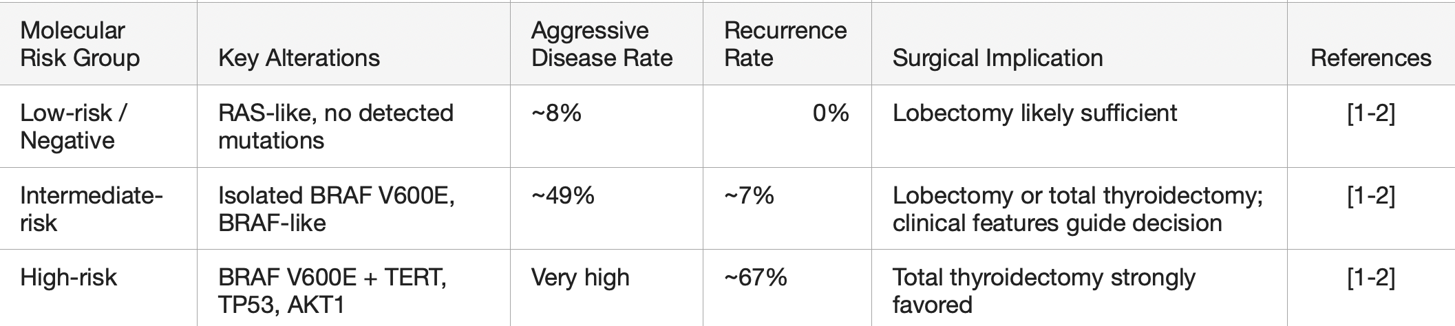
How molecular results may affect decision making for extent of surgery

This page